Ultrasonography

Ultrasonography

Imaging with sound waves is called ultrasound imaging, and is the second most common form of medical imaging in veterinary medicine. When an ultrasound examination is performed, a harmless, high-frequency sound beam – not detectable by humans or pets – is projected into the body of your pet. Ultrasound examinations are complementary to x-rays: they are especially useful in detecting abdominal diseases , reproductive problems and are often able to provide a diagnosis when x-rays cannot. The functioning of various organs and blood flow can be observed to determine if they are malfunctioning.
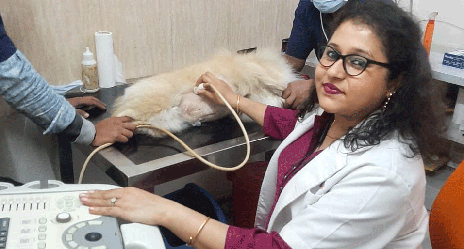
In modern scanning systems sound beam sweeps through the body many times per second. This produces a dynamic, real-time image that changes as the dog ultrasound device moves across a dog’s body. We can use the results of an ultrasound to determine what is ailing your dog, and to devise the most effective treatment protocol.
Common symptoms that may cause a veterinary to use ultrasound include: vomiting, weight loss, Blood in urine, kidney impairment or blockage, Loss of appetite, discharge from vagina, and heart disease.

Have a Question?
Talk to Us!

If you have any questions or would like to learn more about our diagnostic services for companion animals, please fill out our Contact Form or call us directly at 011-66-448-448 for prompt assistance and expert support.

Fill Up the Enquiry Form